
In 1931, electron microscopy took a major step forward. The first prototype electron microscope is generally dated to 1931, but Ruska later described the first electron microscope in the modern sense as being built in 1933; the 1931 work was an early prototype by Max Knoll and Ernst Ruska. This may not seem very impressive at first glance, but it helped overcome one of science’s greatest limits. Visible light has a fixed wavelength range, which meant that some things were simply too small for scientists to study. The invention of the electron microscope did more than just enhance image quality; it made visible structures that had been inaccessible before because of their size.
From light to electrons
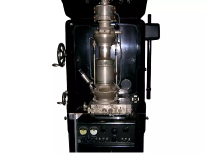
The key concept for the development of the electron microscope originated in an ingenious yet intuitive idea. Scientists assumed electrons could be manipulated in ways similar to light. A small magnetic coil would have a similar effect to a convex glass lens when applied to an electron beam, because the focal point depends on the intensity of the electric current running through the coil.
By 1931, Knoll and Ruska developed the theoretical approach into a functional prototype of the electron microscope. It was a carefully designed device based on the principles of electron optics, intended to surpass conventional optical technology. Light microscopes had their physical limitations, which meant that the resolving power of such a device reached its limit at the shortest wavelength of visible light. Further enhancement of resolution with light microscopes would be impossible, while electrons were much shorter wavelength.

Why 1931 was a watershed for electron microscopy
The chronology of electron optics reveals that 1931 was the precise year when the first electromagnetic microscope was developed. According to the historical review indexed in PubMed, this marked the emergence of electron optics from a pure theoretical concept to reality. The instrument was not created in its final form. Rather, it was conceived as a prototype in which a whole series of theoretical advancements culminated. Therefore, 1931 may be said to have been the time when many technical challenges were simultaneously overcome. These included beam manipulation, magnetic focusing, and the creation of a new type of device.
Its practical applications proved the value of the invention almost immediately, since the microscope could now be subjected to practical tests on real samples. This made it clear that further research would focus on biology and medicine. Three stages from the life cycle of bacteriophage T4 infecting a bacterial cell. Image Credit: Wikimedia Commons
The emergence of a fresh biology perspective
By the end of the 1930s, electron microscopy was being used to investigate bacteria and viruses. The most significant feature of viruses is that they are substantially smaller compared to the objects light microscopes can observe. Early electron micrographs of viruses and phages have shown that the instrument is capable of imaging things which had been impossible before. This diverse usage has been confirmed by a comprehensive scientific article posted in Nature; it has been stated that the application came right after the invention of the microscope in 1931 and is essential for the research of organelles, membranes, pathogenic agents, and tissue ultrastructure.
This opened a fresh biological perspective. After scientists gained the ability to examine small pathogenic agents and tissues in detail, classification became more precise.
Materials Science: The unseen world
Electron microscopy was not confined to the study of living organisms. The 1931 prototype also laid the groundwork for analyzing crystals and nanodevices, helping materials science move from general observation to structural analysis.
Just as it helped scientists study the structure of life, electron microscopy also helped explain why some metals failed and how crystals formed. By combining electromagnetic lenses into an effective device, Knoll and Ruska pushed scientific observation beyond what the naked eye can see and into an unseen world.