Groundbreaking Study Uncovers Mechanism Behind Black Fungus Tissue Destruction
Doctors have identified severe oxidative stress within infected tissues as the primary driver behind the rapid and extensive tissue destruction characteristic of black fungus disease, medically known as mucormycosis. This fungal infection, which saw a dramatic surge during the COVID-19 pandemic, has been particularly devastating due to its aggressive progression.
Understanding Oxidative Stress and Its Role
Oxidative stress occurs when harmful molecules called free radicals accumulate beyond the body's natural antioxidant defenses, leading to significant cellular damage. In the context of mucormycosis, this biochemical imbalance creates an environment where tissues are rapidly destroyed, complicating treatment and recovery.
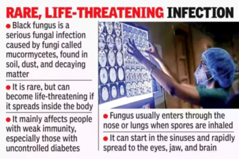
Black fungus is a rare but frequently fatal fungal infection that predominantly targets individuals with compromised immune systems. India experienced a sharp increase in cases during 2021, largely attributed to the widespread use of steroids in COVID-19 treatment and the high prevalence of uncontrolled diabetes among patients.

The Deadly Progression of Mucormycosis
The most dangerous form of the infection typically originates in the sinuses and can swiftly spread to the eyes and brain. Once the infection reaches the brain, mortality rates can escalate to as high as 80%. During the 2021 outbreak, over 3,000 deaths in India were directly linked to COVID-19-associated mucormycosis, highlighting the severity of this public health crisis.
Research Methodology and Key Findings
In a comprehensive study published in the journal Medical Mycology, researchers from the LV Prasad Eye Institute (LVPEI) in Hyderabad collaborated with scientists from the University of Alabama in the United States. The team examined eye and orbital tissues surgically removed from 14 patients suffering from severe mucormycosis, including 10 individuals who had contracted COVID-19.
The infected tissues were meticulously compared with healthy control samples, revealing critical insights:
- Approximately 93% of the patients had diabetes, reinforcing the established connection between diabetes and increased susceptibility to mucormycosis.
- COVID-19 viral proteins were detected in 71% of the cases linked to coronavirus infection, including some patients who had previously recovered from COVID-19.
- Extremely elevated levels of oxidative stress were observed in all infected tissues, triggering multiple cell-death pathways that resulted in rapid tissue necrosis.
These pathological changes were consistent across both COVID-19-related and non-COVID-19 cases, indicating that the fungus itself is the primary agent of tissue destruction.
Clinical Implications and Future Treatment Directions
Dr. Sanhita Roy, head of ocular pharmacology research at LVPEI and lead author of the study, emphasized the significance of these findings. "Our research demonstrates that while COVID-19, steroid therapy, and diabetes significantly elevate the risk of mucormycosis infection, the severe tissue damage is predominantly caused by the fungal pathogen itself," she explained.
This discovery opens new avenues for therapeutic development. Future treatments could potentially combine antifungal agents with antioxidants or other compounds that mitigate oxidative stress, thereby reducing tissue damage and improving patient survival rates. Such integrated approaches could transform the management of this deadly infection, offering hope for better outcomes in future outbreaks.
The study underscores the importance of addressing underlying metabolic conditions like diabetes and carefully monitoring steroid use in vulnerable populations to prevent similar health emergencies.