In an extraordinary display of surgical ingenuity, doctors in China have successfully saved a woman's completely severed ear by first grafting it onto her foot to keep the tissue alive, before finally reattaching it to her head months later. This remarkable procedure highlights the advanced capabilities of modern reconstructive microsurgery.
A Workplace Accident and a Surgical Dilemma
The patient, a woman whose identity was not disclosed, suffered a horrific workplace accident in April. The incident not only tore off her ear but also caused severe injuries to her scalp, neck, and face. When she arrived at Shandong Provincial Hospital in Jinan, the medical team faced a critical challenge.
Qiu Shenqiang, the deputy director of the microsurgery unit, explained that initial attempts to repair the scalp using standard methods failed. The damage to the scalp tissue and blood vessels was too extensive. Crucially, the surrounding skull tissue needed time to heal, making an immediate ear reattachment impossible. The doctors needed a way to preserve the severed ear—a complex organ with delicate cartilage and skin—for a future operation.

The Innovative Solution: A Temporary Home on the Foot
Faced with this problem, the hand, foot, and reconstructive microsurgery team devised a novel plan. They decided to graft the severed ear onto the top of the woman's own foot. Dr. Qiu stated this location was chosen because the arteries and veins in the foot were a suitable size match for those in the ear. Furthermore, the skin on the foot is similar in thinness to the skin on the head, which would simplify the final reattachment.
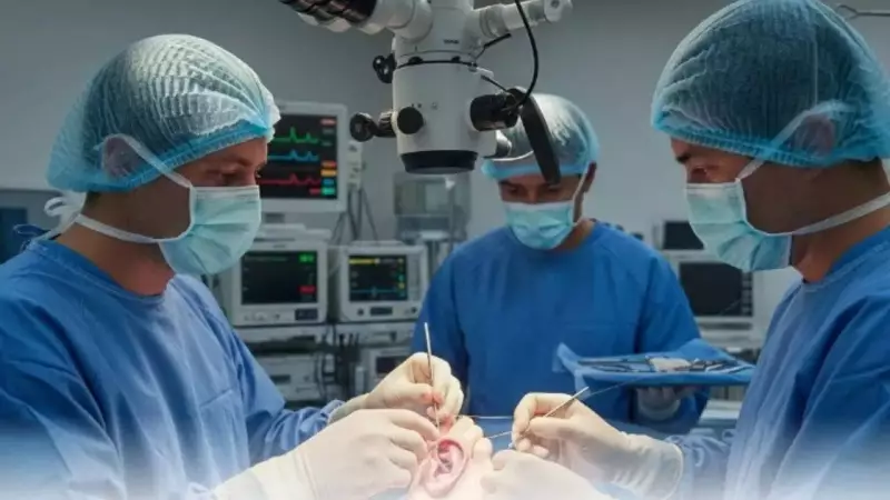
The initial grafting surgery was a marathon effort, lasting 10 hours. One of the most delicate tasks was reconnecting the ear's tiny blood vessels, which were a mere 0.2 to 0.3 millimetres in diameter. The procedure was a success, and the ear began to receive a blood supply from the foot.
Overcoming Crisis and the Final Reattachment
The journey was not without peril. Just five days after the first surgery, doctors observed a dangerous complication: venous reflux. The ear turned a purplish-black colour, indicating poor blood circulation and a high risk of tissue death. To save the graft, the medical team performed an intensive rescue effort. Over the next five days, they carried out manual bloodletting approximately 500 times to relieve pressure and restore proper blood flow.
While monitoring the ear on the foot, surgeons simultaneously worked on reconstructing the woman's scalp. They used skin grafted from her stomach to repair the damaged area on her head. After months of careful recovery and preparation, the team performed the final surgery. The ear, now healthy and viable, was successfully reattached to its original position on the woman's head.
A History of Pioneering Reconstructive Procedures in China
This case is part of a history of innovative reconstructive surgeries performed by Chinese medical teams. In 2013, doctors grew facial tissue on the chest of a burn victim, Xu Jianmei, before transplanting it in a pioneering face transplant. More recently, in 2017, surgeons cultivated an artificial ear on a man's arm for three months before transplanting it to his head after a traffic accident.
These procedures push the boundaries of plastic and reconstructive surgery, offering hope to patients with severe traumatic injuries. The successful ear salvage operation demonstrates how creative thinking and microsurgical expertise can combine to achieve what was once thought impossible, restoring both form and function.