
Pune Hospital Performs Life-Saving Complex Heart Procedure on High-Risk Patient
In a remarkable medical achievement, doctors at a private hospital in Pune have successfully saved the life of a 60-year-old man suffering from renal failure and severe triple vessel disease. The patient's condition was so critical that traditional bypass surgery was deemed too risky due to severely narrowed arteries, making this innovative procedure his only hope.
Critical Condition and Diagnostic Challenges
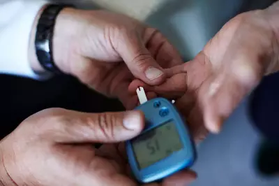
The patient, who was on regular dialysis, experienced dangerous blood pressure drops and irregular heartbeats during dialysis sessions. His condition was rapidly progressing toward complete heart failure, creating an urgent need for intervention. An angiography revealed the severity of his coronary artery disease - all three major heart arteries (left anterior descending, left circumflex, and right coronary artery) had significant plaque buildup, severely restricting blood flow to his heart.
What made this case particularly challenging was the extreme calcification of the patient's blood vessels. The hardened calcium deposits had become nearly impossible to break through conventional means. Compounding the problem, the patient's heart had weakened dramatically, pumping at only 20% of its full capacity. This combination of factors created an exceptionally high-risk scenario for any medical procedure.

Innovative Surgical Solution
Cardiologist Dr. Suraj Ingole, head of the department of cardiac sciences at VishwaRaj Hospital in Loni, Pune, led the medical team that developed a sophisticated solution. They performed a calcium orbital atherectomy followed by high-risk angioplasty to address the patient's complex condition.
"We chose angioplasty considering the patient's condition and the multiple complexities involved," explained Dr. Ingole. "The highly calcified blood vessels, which were extremely hard to break, needed to be cleared before we could deploy stents. Therefore, we conducted a calcium orbital atherectomy, which is a minimally invasive procedure employing a diamond-coated crown that rotates and orbits to cut through the calcium plaque."
The medical team also utilized intravascular ultrasound technology during the procedure. This catheter-based, minimally invasive imaging technique uses high-frequency sound waves to generate detailed, real-time 360-degree cross-sectional images from inside blood vessels, providing crucial guidance for the delicate operation.
Successful Outcome and Recovery
The patient was admitted on February 12, underwent the complex surgery on February 14, and was discharged just two days later on February 16. Following the successful clearance of the calcified vessel, doctors placed a stent in the left anterior descending artery.
"Once the calcified vessel was cleared, we were able to successfully deploy the stent," added Dr. Ingole. "The patient underwent dialysis the very next day without any complications, which was a significant milestone in his recovery."
Medical professionals plan to perform another angioplasty in one to two months to address the remaining affected vessels. The anesthetist present during the procedure emphasized the case's complexity, noting that "the affordable nature of this process represents a new ray of hope for common people in the eastern periphery of Pune, where such advanced procedures are typically rare."
This successful intervention demonstrates how innovative medical techniques can overcome even the most challenging cardiovascular cases, particularly when combined with expert medical judgment and advanced technology.