
New imaging technology has brought unprecedented clarity to ancient Egyptian mummified body parts that remained sealed for over two millennia. Scientists utilized advanced CT and 3D scanning techniques to examine limbs, skulls, and a foot dating back more than 2,300 years.
Details of the Scanned Remains
The remains, believed to be from between 401 and 259 BCE, have been preserved in museum collections for decades but had never been studied in such detail. The scans reveal a clearer internal view of bones, bandages, and structural damage that was previously hidden. Experts note that even small features, such as missing bone sections and traces of disease, are now visible without unwrapping or damaging the mummies. This rare glimpse into ancient preservation methods, which are still not fully understood, offers valuable insights.
The CT scan focused on multiple body parts, including two skulls, two lower limbs, a hand, and a single foot wrapped tightly in linen bandages. Each item was scanned using high-resolution imaging systems, yielding sharper results than earlier attempts. Researchers report that previous examinations missed several internal details.

The Foot and Its Mysteries
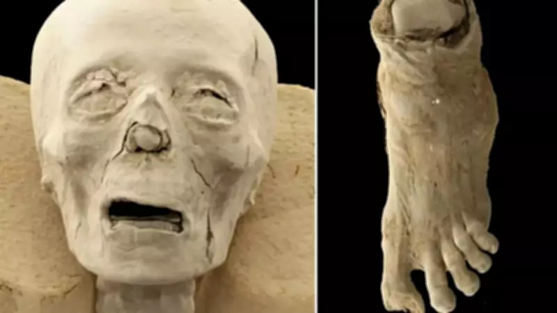
One of the most striking findings is the foot, which still retains its wrappings. The bones inside are clearly visible through the scans, and a portion of the big toe appears missing. This damage may have occurred either before or after mummification, but the exact cause remains uncertain. Interestingly, the foot was once thought to belong to a bird, but that idea has now been ruled out.
Signs of Disease and Age Variation
Some of the bones show signs of disease. One lower limb appears affected by osteoporosis, a condition that weakens bone structure and increases fracture risk. In ancient times, this could have caused serious mobility problems or even fatal injuries. Another limb seems to belong to a younger individual, with less developed bones and growth patterns suggesting a different age group. The hand remains more ambiguous; researchers are still determining whether it belonged to a child or an adult, analyzing bone structure and development markers.
Early findings indicate multiple individuals rather than a single burial set, though nothing is confirmed completely yet.
Imaging Methodology
The imaging work was conducted at a medical research center affiliated with Semmelweis University. Scientists used CT scans and 3D reconstruction methods to see inside the wrappings without cutting or unwrapping. The images reveal layered bandages pressed tightly around bones. Skulls appear structurally intact, though slightly deformed due to time and pressure, with some facial bone outlines and teeth positions still visible. A few scans even show internal gaps between bandage layers, which may have been intentionally created during mummification, though experts continue to debate this.
Ancient Preservation Techniques
Ancient Egyptian mummification remains one of history's most complex preservation practices. Bodies were treated with drying agents and wrapped carefully in linen, likely for preservation in the afterlife. Some researchers believe specific materials were used to slow decay, while others suggest ritual methods played a role. The latest scans raise more questions than answers, revealing careful wrapping but also variation between individuals.