
A young man in his late 20s endured five years of recurring seizures and growing uncertainty. Initially, a small lesion in his brain was believed to be tuberculosis, a common suspicion for such MRI findings. He underwent 18 months of anti-tubercular treatment, hoping for improvement.
Treatment Failure and Worsening Symptoms
Instead of responding to therapy, the lesion continued to enlarge. His seizures persisted, and his symptoms worsened, raising concerns for him and his family. The unusual behavior of the lesion prompted doctors to question whether it was truly brain tuberculosis or a rare condition.
Surgical Intervention with Advanced Technology
As the lesion grew despite medical treatment, doctors at Apollo Hospitals, Sheshadripuram, decided surgery was necessary for both treatment and diagnosis. The neurosurgical team used advanced neuro-navigation technology to precisely locate the abnormal area, minimize the craniotomy size, and reduce surgical risk.

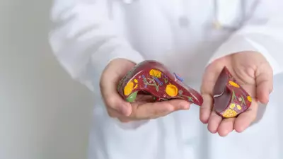
Histopathological Diagnosis
The excised lesion was examined and confirmed to be intracranial sparganosis, a rare parasitic worm infection of the brain. After diagnosis, the patient received appropriate anti-helminthic therapy and is recovering well.
Dr. Adesh J, Senior Consultant in Neurosurgery & Spine Surgery at Apollo Hospitals, said, "When a brain lesion continues to enlarge despite adequate treatment, it becomes important to reconsider the diagnosis. In this case, surgery was planned not only to remove the lesion safely but also to obtain tissue confirmation."
Rapid Recovery and Discharge
The surgery was successful. The patient regained consciousness soon after, started oral intake the same day, and walked on the first postoperative day. He was discharged within 48 hours, bringing relief after years of uncertainty.
Dr. Raj Kumar Pannem, Consultant in Neurosurgery, added, "Sparganosis of the brain is extremely rare and can mimic common conditions like tuberculosis on imaging. This case highlights why tissue diagnosis is important when a lesion does not respond as expected. Neuro-navigation helped localize the lesion accurately, minimize surgical morbidity, and support faster recovery."
This case underscores the importance of considering rare causes when a presumed diagnosis does not follow the expected course. Today, after years of seizures and uncertainty, the patient is recovering well, with doctors monitoring his progress.