A 44-year-old woman arrived at a Pune hospital with severe pain on the left side of her abdomen. During evaluation, doctors discovered an unexpected anatomical finding: her internal organs were a complete mirror image of normal anatomy, a condition called Situs Inversus Totalis (SIT). This rare congenital variant occurs in roughly 1 in 25,000 people and can complicate diagnosis, as symptoms often present on the opposite side from what clinicians expect.
Challenges in Diagnosis and Surgery
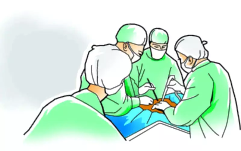
SIT also presents technical challenges during surgery. Standard incision sites and operative approaches may need to be adapted, and surgical teams must plan carefully to avoid confusion. In this particular case, the surgical team successfully adjusted their strategy and completed the operation without complications.
Dr. Abhijit Whatkar, senior robotic and laparoscopic surgeon at Noble Hospitals and Research Centre, explained: “The patient presented with complaints of severe abdominal pain for four days prior to admission in the first week of May. Her complicated medical history — hypertension, paraumbilical incisional hernia (bulge at the site of previous surgical scar) for 15 years, tubectomy, long-standing HIV positive status, and a history of tuberculosis — made the surgery challenging.”

He added, “In addition, her mirror anatomy added to the challenge. The woman’s liver and gallbladder, which are usually to the right, were on the left, and she had dextrocardia, where the heart’s apex is pointed left instead of the usual right.”
Diagnostic Findings
On the day of admission, ultrasonography and magnetic resonance cholangiopancreatography (MRCP) revealed a condition called cholelithiasis, hardened deposits in the gallbladder, along with SIT. The patient was planned for laparoscopic cholecystectomy with concurrent Mayo’s repair for paraumbilical hernia.
“During the surgery, we first treated the hernia near the belly button by pushing back the trapped fatty tissue into the abdomen. A camera port was then inserted through the same opening, and a few more ports were made in different areas of the abdomen for laparoscopic surgery according to mirror image ergonomics,” Dr. Whatkar said.
Surgical Adaptations
Since the organs were reversed, the surgeon performed the procedure in a “mirror-image” manner while standing on the patient’s right side. A special dye imaging technique was used to clearly identify the gallbladder ducts and blood vessels before they were clipped and cut safely.
Dr. Divij Mane, deputy managing director at Noble Hospitals, emphasized that treating rare conditions with other complex pre-existing conditions requires meticulous operative planning and ergonomic modifications. He stated, “Laparoscopic cholecystectomy is considered the gold standard treatment for symptomatic cholelithiasis. However, performing the procedure in patients with situs inversus totalis requires modification of standard operative strategies, trocar placement, and surgeon ergonomics. The gallbladder was then removed through keyhole surgery. Bleeding was checked and controlled, and a temporary drainage tube was placed inside the abdomen to remove excess fluid after surgery. Finally, all small cuts were closed, and the abdominal wall defect from the hernia was repaired.”
Recovery and Follow-Up
The patient was operated on in the first week of May and was discharged in just three days. During her last follow-up on June 3, she had completely recovered, and the hernia sutures had healed, indicating no further complications.